Mitosis and Its Stages | Phases of Mitosis
Channel: Dr. Najeeb Lectures
Category: Education
Tags: steps of mitosismedical educationsister chromatidsmitosis & its stagesusmle step 1dna replicationcellninja nerdmitosis stagesdr najeeb lecturesdna replication animationmitosis cell divisiondnainterphasemitosis vs meiosisamoeba sistersdr najeebphases of mitosisprophasebiologycell cyclegenescytokinesischromosomescell divisionplabmitosis and its stagesmitosisusmlespindlescellscell biologycell multiplicationdna duplication
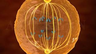
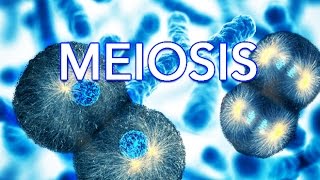
Description: Like this video? Sign up now on our website at DrNajeebLectures.com to access 800+ Exclusive videos on Basic Medical Sciences & Clinical Medicine. These are premium videos (NOT FROM YOUTUBE). All these videos come with English subtitles & download options. Sign up now! Get Premium Access starting from $99 ONLY! #Mitosis #Mitosis&CellDivision #CellDivision #PhasesofMitosis In cell biology, mitosis is a part of the cell cycle in which replicated chromosomes are separated into two new nuclei. Cell division gives rise to genetically identical cells in which the total number of chromosomes is maintained. Therefore, mitosis is also known as equational division. In general, mitosis (division of the nucleus) is preceded by the S stage of interphase (during which the DNA is replicated) and is often followed by telophase and cytokinesis; which divides the cytoplasm, organelles and cell membrane of one cell into two new cells containing roughly equal shares of these cellular components. The different stages of Mitosis altogether define the mitotic (M) phase of an animal cell cycle—the division of the mother cell into two daughter cells genetically identical to each other. The process of mitosis is divided into stages corresponding to the completion of one set of activities and the start of the next. These stages are prophase, prometaphase, metaphase, anaphase, and telophase. During mitosis, the chromosomes, which have already duplicated, condense and attach to spindle fibers that pull one copy of each chromosome to opposite sides of the cell. The result is two genetically identical daughter nuclei. The rest of the cell may then continue to divide by cytokinesis to produce two daughter cells. The different phases of mitosis can be visualized in real time, using live cell imaging. Producing three or more daughter cells instead of the normal two is a mitotic error called tripolar mitosis or multipolar mitosis (direct cell triplication / multiplication). Other errors during mitosis can induce apoptosis (programmed cell death) or cause mutations. Certain types of cancer can arise from such mutations. Mitosis occurs only in eukaryotic cells. Prokaryotic cells, which lack a nucleus, divide by a different process called binary fission[citation needed]. Mitosis varies between organisms. For example, animal cells undergo an "open" mitosis, where the nuclear envelope breaks down before the chromosomes separate, whereas fungi undergo a "closed" mitosis, where chromosomes divide within an intact cell nucleus. Most animal cells undergo a shape change, known as mitotic cell rounding, to adopt a near spherical morphology at the start of mitosis. Most human cells are produced by mitotic cell division. Important exceptions include the gametes – sperm and egg cells – which are produced by meiosis. There are five phases of mitosis. Each phase is used to describe what kind of change the cell is going through. The phases are prophase, prometaphase, metaphase, anaphase and telophase. Prophase: During prophase, chromatin (tangled-up DNA) in the nucleus condense into chromosomes (bunched-up DNA). Pairs of centrioles move to opposite sides of the nucleus. Spindle fibers begin to form a bridge between the ends of the cell. Prometaphase: During prometaphase, the nuclear envelope around the chromosomes breaks down. Now there is no nucleus and the sister chromatids are free. A protein called a kinetochore forms at each centromere. Long thin proteins reach across from opposite poles of the cell and attach to each kinetochore. Metaphase: During metaphase, the pair of chromatids are aligned by the pushing and pulling of the attached kinetochore microtubules, similar to a game of "tug of war". Both sister chromatids stay attached to each other at the centromere. The chromosomes line up on the cell's equator, or center line, and are prepared for division. This is the longest phase of mitosis. Anaphase: During anaphase, the sister chromatids split apart and move from the cell's equator (metaphase plate) to the poles of the cell. The kinetochore is attached to the centromere. The microtubules hold on to kinetochore and shorten in length. Another group of microtubules, the non-kinetochore microtubules, do the opposite. They become longer. The cell begins to stretch out as the opposite ends are pushed apart. Telophase: Telophase is the final stage in mitosis: the cell itself is ready to divide. One set of chromosomes is now at each pole of the cell. Each set is identical. The spindle fibers begin to disappear, and a nuclear membrane forms around each set of chromosomes. Also, a nucleolus appears within each new nucleus and single stranded chromosomes uncoil into invisible strands of chromatin.